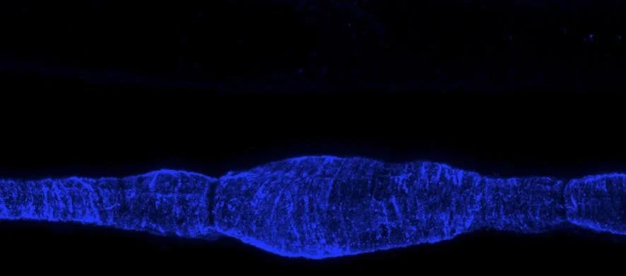
Lymphatic muscle cells in the wall of a rat mesenteric lymphatic vessel. The bulge at the centre of the image indicates the location of a lymphatic valve. From Zawieja et al., Am J Physiol Heart Circ Physiol 302:H643-H653, 2012.
Lymphatic Muscle Contraction Dynamics
Contraction of lymphatic muscle cells is crucial for propelling lymph through the body. These cells are a hybrid of cardiac, smooth and skeletal muscle types. Experimental evidence on whole cannulated vessels shows that the acto-myosin complex in these individual cells facilitates phasic contractions which in produce most of the pumping action. The smooth muscle components regulate the diameter, and thus the resistance to flow from adjacent vessels. The contractile signature of lymphatics has not been studied at the single-cell level. We aim to induce active contractions in single isolated human lymphatic muscle cells via a custom-built bioreactor and subsequently measure their biomechanics on a substrate through traction force microscopy.

The Faciliflow implant
Lymph Node Implant
Around 1 in 5 breast cancer patients will develop lymphoedema in the arm after surgery involving removal of lymph nodes. Lymphoedema is a chronic, incurable condition which causes localised swelling of tissue as a result of abnormal lymphatic drainage. It is a debilitating condition that can cause significant physiological and psychological trauma. The device under development will be implanted in place of surgically removed nodes to restore lymph flow and hopefully prevent the formation of lymphoedema. The implant contains a hydrogel encapsulating the lymphangiogenic growth factor VEGF-C, which is released by two time-dependent mechanisms. The combination of the release profiles provide a prolonged release of VEGF-C, encouraging lymphatic vessels to reconnect.

Device for Modulating Lymphatic Pumping
The current standard of care for lymphoedema is complex decongestive therapy (CDT), which is a combination of compression therapy, manual lymphatic drainage (MLD), exercise, and skin care. Using our validated computational models of lymphatic pumping, we discovered that an external pressure below the pressure inside of the lymph vessel (or a positive transmural pressure) maximises pumping. External compression, on the other hand, restricts re-filling of the vessel after contraction, which practically shuts down pumping. This finding led to the development of a new device that uses a cyclic negative pressure to maximize lymph drainage. It is currently under trials with human volunteers.
Lymphoedema in the arm of a breast cancer patient. Image downloaded 26 April 2019 from https://lymphaticsolutions.com.au/early-detection-for-breast-cancer-related-lymphedema/

Microfluidic chamber used to study cell migration in response to chemokine gradients.
Immune cell migration dynamics in response to chemokine concentration gradients
All inflammatory processes require precise positioning and migration of immune cells to fight pathogens efficiently and be resolved once the pathogen threat is eliminated. Immune cells constantly follow trails of attractant molecules called chemokines to exchange information about antigens and trigger appropriate reactions. Little is known quantitatively about how extra-cellular chemokine distribution is controlled. We use numerical modelling and in-vitro microfluidic experiments in synergy to reproduce, isolate, and quantify the biophysical and biochemical phenomena known to affect it in-vivo. We aim to apply this to better understand inflammation and inflammatory diseases, including atherosclerosis.

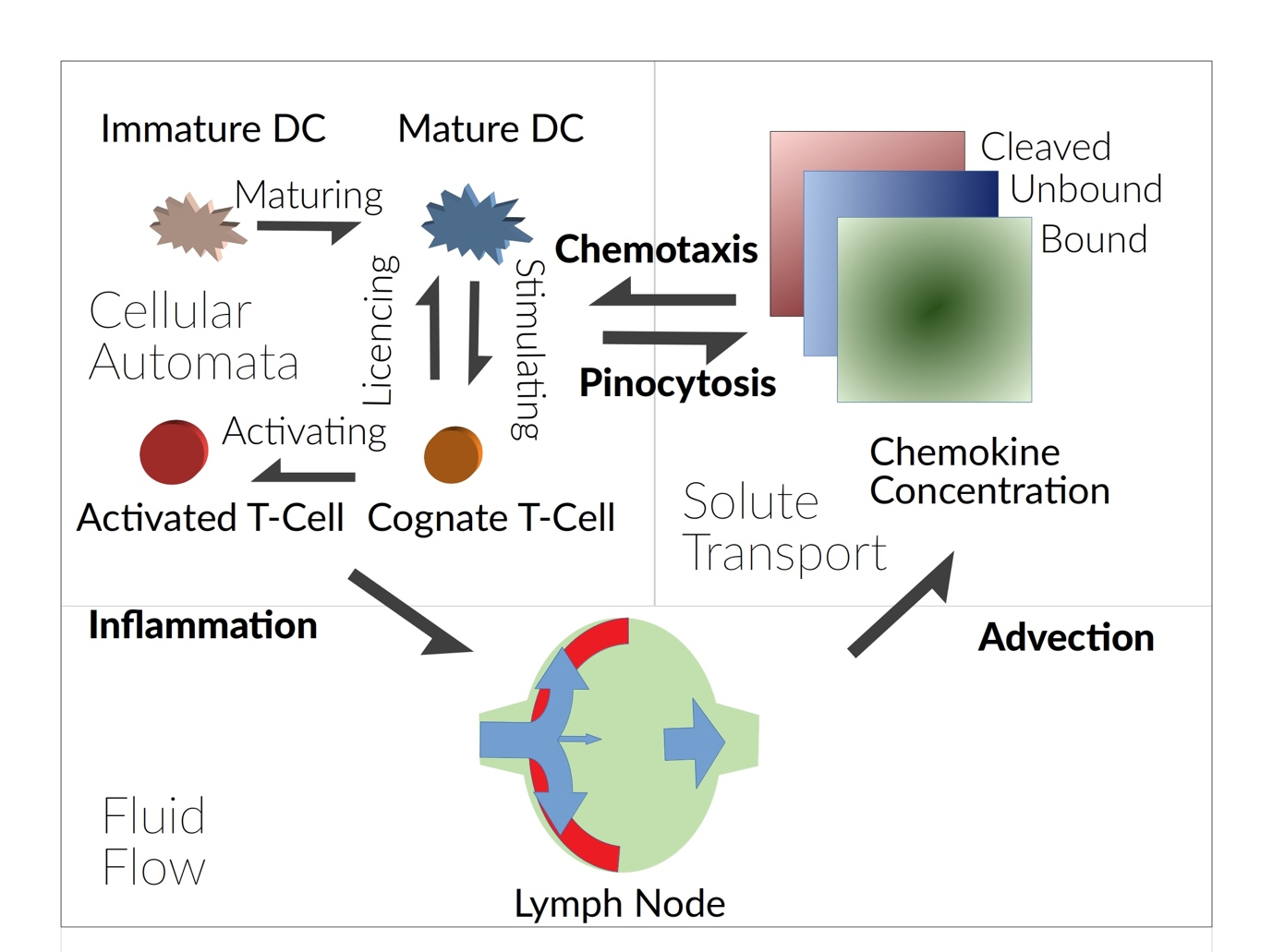
Illustration of how we model immune cell interactions in lymph nodes using Agent-Based Modelling.
Lymphatic System Mass Transport
The lymphatic system has multiple functions; it is a drainage system that allows maintenance of fluid balance throughout the body, a crucial component of the immune system, and a provides a major route for cancer cell metastasis. [1]. Lymph nodes are stationed along the lymphatic network, sample the draining lymph and allow initiation of the adaptive immune response if antigen is detected. By using computer models we can see beyond experiments and understand the complex interactions between our cells, tissues and fluids. By creating and refining a set of rules for how T-cells and dendritic cells behave we can create simulations of hundreds of thousands of cells called cellular automata. This allows us to explore how the immune cells interact with each other to protect us from infection [1].
By studying small signalling proteins, called chemokines and how they build up in the lymph nodes we can better understand how cell motion and function is coordinated [2]. How this helps our immune system when we are healthy and what can go wrong in disease.
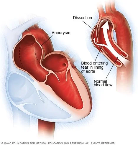
Ascending Aortic Dissection
An aortic dissection is a serious condition caused by the tear occurring between the innermost and middle layers of the aorta. The dissections that occur on the ascending part of the aorta are known as Type A dissections whereas Type B dissections occur in the descending portion of the aorta. As blood surges through the tear, it causes the inner and middle layers of the aorta to separate (dissect). People who have aortic aneurysms are at a higher risk of dissection influenced by the size of the enlargement, prior medical conditions and heredity. Complicated aortic dissections include those that rupture or have malperfusion (abnormal perfusion of blood) to other organs. Biomechanical factors have been found to play a major role in the development and propagation of aortic dissection. The emergence of advanced imaging techniques have led to multiple new findings in the areas of thoracic aortic aneurysm formation and aortic prosthesis performance assessment. Here in Moore’s Lab, we are analysing the disease through a multi-disciplinary approach that innovatively combine the state of the art research techniques (mechanical characterisation of diseased tissue, computational imaging, modelling and bioinformatics) to understand the variable progression of the condition and thereby develop improved patient-specific treatment modalities..

Illustration of stem cells
Cell Therapy Injection Strategies
Stem cells have shown promise for treatment of multiple diseases. Unfortunately, the techniques used to inject cells apply large forces to the cells that rupture their membranes, killing the cells. We are developing a combination of suspension media and injection devices that minimise cell death.
Title
The lymphatic system has multiple functions; it is a drainage system that allows maintenance of fluid balance throughout the body, a crucial component of the immune system, and a provides a major route for cancer cell metastasis. [1]. Lymph nodes are stationed along the lymphatic network, sample the draining lymph and allow initiation of the adaptive immune response if antigen is detected. By using computer models we can see beyond experiments and understand the complex interactions between our cells, tissues and fluids. By creating and refining a set of rules for how T-cells and dendritic cells behave we can create simulations of hundreds of thousands of cells called cellular automata. Which allows to explore how they interact with each other to protect us from infection [1].
By studying small signalling proteins, called chemokines and how they build up in the lymph nodes we can better understand how cell motion and function is coordinated [2], how this helps our immune system when we are healthy and what can go wrong in disease.